Search Again
We support boolean queries, use +,-,<,>,~,* to alter the weighting of terms
ConDeTri
Software tool as content dependent read trimmer for Illumina data. Content dependent read trimming software for Illumina/Solexa sequencing data.
- Resource
- SciCrunch
- 13 years ago - by Anonymous
Comprehensive Perl Archive Network
Welcome to CPAN, where you you will find All Things Perl. CPAN is the Comprehensive Perl Archive Network, a large collection of Perl software and documentation. You can begin exploring from either http://www.cpan.org/, http://www.perl.com/CPAN/ or any of the mirrors listed at http://www.cpan.org/SITES.html. Note that CPAN is also the name of a Perl module, CPAN.pm, which is used to download and install Perl software from the CPAN archive. This FAQ covers only a little about the CPAN module and you may find the documentation for it by using perldoc CPAN via the command line or on the web at http://search.cpan.org/dist/CPAN/lib/CPAN.pm. Sponsors: CPAN works with the generosity and cooperation of hundreds of developers, over 100 participating mirrors, funet.fi donating the network bandwidth, storage space and computing power, volunteers who help keep everything together and users whose interest in Perl keep the archive alive and growing. Keywords: Comprehension, Perl, Archive, Software, Documentation,
- Resource
- SciCrunch
- 17 years ago - by Anonymous
Grantees Meeting for NITRC
This project is meant for planning the NITRC Grantee meetings. A website for organizing meetings for the Neuroimaging Informatics Tools and Resources Clearinghouse, to facilitate participants meeting one another, and promote discussion of common interests and collaboration.
- Resource
- SciCrunch
- 11 years ago - submitted by Kristen Jensen
Gemma
Resource for reuse, sharing and meta-analysis of expression profiling data. Database and set of tools for meta analysis, reuse and sharing of genomics data. Targeted at analysis of gene expression profiles. Users can search, access and visualize coexpression and differential expression results.
- Resource
- SciCrunch
- 17 years ago - by Anonymous
Protein Data Bank Bind Database
A database of binding affinities for the protein-ligand complexes in the Protein Data Bank (PDB). The PDBbind database is a collection of the experimentally measured binding affinities exclusively for the protein-ligand complexes available in the Protein Data Bank (PDB). It thus provides a link between energetic and structural information of those complexes and may be of great value to various molecular recognition studies. This site was last updated in 2007. The updated version of the resource is maintained by the Shanghai Institute of Organic Chemistry (http://www.pdbbind.org.cn).
- Resource
- SciCrunch
- 17 years ago - by Anonymous
dcm2nii
A tool for converting images from the complicated formats used by scanner manufacturers (DICOM, PAR/REC) to the NIfTI format used by various scientific tools. dcm2nii works for all modalities (CT, MRI, PET, SPECT) and sequence types.
- Resource
- SciCrunch
- 11 years ago - submitted by Kristen Jensen
Matlab Health and Socio-Economic Survey
A data set of the health and socioeconomic factors that affect the elderly in Matlab, a region of rural Bangladesh. The survey captures measurements and statistics such as adult survival, health status, health care utilization, resource flows between generations and the impact of community services and infrastructure on adult health care. Data was collected through surveys that touch on four topics: household and individual information; determinants of natural fertility; migration out of the community; and community and provider survey of healthcare and education infrastructure.
- Resource
- SciCrunch
- 15 years ago - by Anonymous
GIMME
Software Matlab toolbox for directed functional connectivity analysis of fMRI BOLD signal from predefined regions of interest. It recovers true structure of connections and estimates weights attributed to each connection. Obtains patterns at group and individual levels.
- Resource
- SciCrunch
- 11 years ago - submitted by Kristen Jensen
Pychopper
Software tool to identify, orient and trim full length Nanopore cDNA reads. Able to rescue fused reads.
- Resource
- RRID-Legacy
- 6 years ago - submitted by John Hamilton
JDC Computer Resource
Implements and maintains specialized computer facilities required by the DERC investigators. A major emphasis of JoslinResearch.org is to provide dynamic web applications that serve essential informational needs of the research community at the Joslin Diabetes Center. These applications provide such functions as automating media provisioning services to DERC laboratories, distributing DERC event schedule information, providing access to troubleshooting information, fully-searchable call schedules with contact information, text messaging services, and access to information about Section, Investigator and Fellow projects and affiliations. All applications adhere to the JoslinResearch.org paradigm of distributed administration without requiring knowledge of web design or web programming. For DERC use only.
- Resource
- SciCrunch
- 13 years ago - by Anonymous
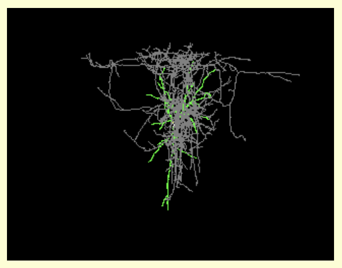
L-Neuron
A program which creates anatomically realistic virtual neurons using the formalism of the Lyndenmayer systems to implement sets of neuroanatomical rules discovered by several research groups. The program algorithms read in experimental data - in the form of statistical distributions - to generate virtual structures. L-Neuron samples the values of the parameters within these statistical distributions in a stochastic (random) fashion during dendritic growth.
- Resource
- SciCrunch
- 11 years ago - submitted by Kristen Jensen
TransDecoder
Software tool to identify candidate coding regions within transcript sequences, such as those generated by de novo RNA-Seq transcript assembly using Trinity, or constructed based on RNA-Seq alignments to genome using Tophat and Cufflinks.Starts from FASTA or GFF file. Can scan and retain open reading frames (ORFs) for homology to known proteins by using BlastP or Pfam search and incorporate results into obtained selection. Predictions can then be visualized by using genome browser such as IGV.
- Resource
- SciCrunch
- 7 years ago - submitted by Shuang Jiang
Arthritis research campaign
http://www.arc.org.uk/
- Resource
- SciCrunch
- 17 years ago - submitted by Timothy Tsui
Neuron-C
A simulation language for modeling biophysically realistic neural circuits (1 to 10,000 neurons) and simulating physiology experiments on it. Programs for plotting and displaying data are included.
- Resource
- SciCrunch
- 11 years ago - submitted by Kristen Jensen
CoVEffect's Dataset: Effects of SARS-CoV-2 Mutations and Variants
Supporting data for "CoVEffect: Interactive System for Mining the Effects of SARS-CoV-2 Mutations and Variants Based on Deep Learning". This dataset include both manually curated and machine-mined annotations of abstracts on SARS-CoV-2 mutations and variants' effects. The abstracts are a subset of the COVID-19 Open Research Dataset.
- Resource
- SciCrunch
- 3 years ago - submitted by Francesco Invernici
Allen Mouse Brain Reference Atlas
Allen Mouse Brain Atlas includes full color, high resolution anatomic reference atlas accompanied by systematic, hierarchically organized taxonomy of mouse brain structures. Enables interactive online exploration of atlas and to provide deeper level of 3D annotation for informatics analysis and viewing in Brain Explorer 3D viewer.
- Resource
- SciCrunch
- 16 years ago - by Anonymous
Ruby NIfTI
A library for handling NIfTI data in the Ruby programming language. Ruby NIfTI supports basic read and write access to NIfTI files, including basic and extended header information and image information. It doesn't attempt to touch the image data but it does provide access to qform and sform orientation matrices. It also provides a nice interface to get at NIfTI info from within Ruby.
- Resource
- SciCrunch
- 11 years ago - submitted by Kristen Jensen
Rhode Island Hospital Molecular Pathology Core Facility
Core of COBRE Center at department of Pathology located on sixth floor of Aldrich building at Rhode Island Hospital.Provides services, instrumentation, training and support. Equipment includes ArcturusXT laser capture microdissector, Stratagene MX3005p realtime PCR instrument, BioRad iCycler, Agilent Bioanalyzer, Olympus fluorescence-capable microscope with image analysis software, Ventana Discovery immunohistochemistry processor, microtome, cryostat, Beecher tissue arrayer and Aperio ScanScope.Tissue bank has 40 cu ft of ultra-low freezer space and provides human tumor tissue samples to all interested investigators who have received IRB approval for academic testing and research.Services include Tissue Sample Processing,Histology,Immunohistochemistry,Digital slide scanning by Aperio ScanScope,Tissue Microarrays,DNA/RNA extraction, Microdissection,Macrodissection verification by pathologist,Digital image analysis,Visium Spatial Gene Expression Assay,RNAscope Assay,Enzyme Linked Immunosorbent Assays (ELISAs),Technical assistance,Pathologist consultation,Technical assistance, Equipment use and training.
- Resource
- SciCrunch
- 3 years ago - submitted by Edyta Vieth
Institute for Magnetic Resonance Safety, Education and Research
The Institute for Magnetic Resonance Safety, Education, and Research (IMRSER) was formed in response to the growing need for information and research on matters pertaining to magnetic resonance (MR) safety. The IMRSER is the first independent, multidisciplinary, professional organization devoted to promoting awareness, understanding, and communication of MR safety issues through education and research. Mission Statement To promote awareness and understanding of MR safety, To disseminate information regarding current and emerging MR safety issues, To develop and provide materials and resources to facilitate MR safety-related education and training, To respond to critical MR safety issues with a sense of urgency, and To advance the field of MR safety through support of scientific research. Functions and activities of the IMRSER include development of up-to-date MR safety materials and dissemination of this information to the MR community. This is accomplished predominantly through the efforts of the Advisory Boards. Members of the Advisory Boards of the Institute for Magnetic Resonance Safety, Education, and Research (IMRSER) are charged with creating recommendations, guidelines, position papers, and educational materials pertaining to existing or emerging MR safety issues. This is achieved by utilizing the pertinent peer-reviewed literature and by relying on each members extensive clinical, research, or other appropriate experience. Notably, documents developed by the IMRSER incorporate MR safety guidelines and recommendations created by the International Society for Magnetic Resonance in Medicine (ISMRM), the American College of Radiology (ACR), the Food and Drug Administration (FDA), the National Electrical Manufacturers Association (NEMA), the Medical Devices Agency (MDA), the International Electrotechnical Commission (IEC), and other similar organizations. The IMRSERs rigorous development and review process for MR safety documents ensures that authoritative and relevant information is produced in a timely manner for rapid dissemination to the MR community. The MR safety information is provided to MR healthcare professionals and others as hard copy and electronic publications. Additionally, this information is posted on the IMRSER web site as well as on www.MRIsafety.com (currently with over 92,000 registered users). The Institute for Magnetic Resonance Safety, Education, and Research permits all members of the MR community to use the MRI Safety Guidelines posted on this web site. Please be sure to read and understand our disclaimer.
- Resource
- SciCrunch
- 17 years ago - by Anonymous
About
The SciCrunch Infrastructure was developed as a cooperative data platform to be used by diverse communities in making data more FAIR.
Contact Us
 scicrunch.org