Reprogramming reactive glia into interneurons reduces chronic seizure activity in a mouse model of mesial temporal lobe epilepsy.
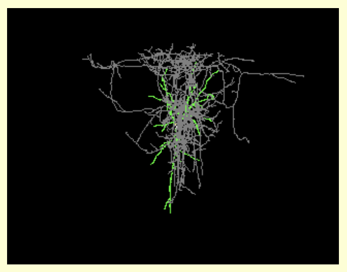
Reprogramming brain-resident glial cells into clinically relevant induced neurons (iNs) is an emerging strategy toward replacing lost neurons and restoring lost brain functions. A fundamental question is now whether iNs can promote functional recovery in pathological contexts. We addressed this question in the context of therapy-resistant mesial temporal lobe epilepsy (MTLE), which is associated with hippocampal seizures and degeneration of hippocampal GABAergic interneurons. Using a MTLE mouse model, we show that retrovirus-driven expression of Ascl1 and Dlx2 in reactive hippocampal glia in situ, or in cortical astroglia grafted in the epileptic hippocampus, causes efficient reprogramming into iNs exhibiting hallmarks of interneurons. These induced interneurons functionally integrate into epileptic networks and establish GABAergic synapses onto dentate granule cells. MTLE mice with GABAergic iNs show a significant reduction in both the number and cumulative duration of spontaneous recurrent hippocampal seizures. Thus glia-to-neuron reprogramming is a potential disease-modifying strategy to reduce seizures in therapy-resistant epilepsy.
Pubmed ID: 34592167 RIS Download
Research resources used in this publication
- Leica TCS SP5 II microscope (RRID:SCR_018714) (instrument resource)
- Leica TCS SPE (RRID:SCR_002140) (instrument resource)
- Zeiss LSM 710 Confocal Inverted Microscope (RRID:SCR_018063) (instrument resource)
- Neurolucida 360 (RRID:SCR_016788) (software resource)
- ImageJ (RRID:SCR_003070) (software resource)
- Black Zen software (RRID:SCR_018163) (software resource)
- C57BL/6J (RRID:IMSR_JAX:000664) (organism)
Additional research tools detected in this publication
Antibodies used in this publication
- VIP (vasoactive intestinal polypeptide) (RRID:AB_572270)
- Anti-VGAT (RRID:AB_887869)
- RFP Antibody Pre-adsorbed (RRID:AB_2209751)
- Rabbit Anti-Somatostatin Antibody, Unconjugated (RRID:AB_518614)
- ANTI-OLIG-2 (RRID:AB_570666)
- Anti-Parvalbumin (RRID:AB_2156476)
- Anti-Neuropeptide Y (RRID:AB_2721083)
- Rabbit Anti-NeuN (polyclonal) Polyclonal Antibody, Unconjugated (RRID:AB_10807945)
- Anti-NeuN (RRID:AB_2619988)
- anti-mCherry (RRID:AB_2333095)
- Anti-MAP 2 (RRID:AB_2619881)
- Anti Iba1, Rabbit antibody (RRID:AB_839504)
- Glial Fibrillary Acidic Protein (Multipurpose) (RRID:AB_10013382)
- Anti-Gephyrin (RRID:AB_2651176)
- GAD67 antibody [K-87] - Neuronal Marker (RRID:AB_448990)
- GFAP Monoclonal Antibody (131-17719) (RRID:AB_2535827)
- Anti-GFP antibody (RRID:AB_300798)
- Guinea pig Anti-Doublecortin , Unconjugated (RRID:AB_1586992)
- Mouse Anti-Calretinin Monoclonal antibody, Unconjugated (RRID:AB_94259)
- Anti-c-Fos (RRID:AB_2864765)
- Rat Anti-BrdU Monoclonal Antibody, Unconjugated, Clone BU1 / 75 (ICR1) (RRID:AB_305426)
Associated grants
- Agency: Wellcome Trust, United Kingdom
- Agency: Medical Research Council, United Kingdom
Id: MR/N026063/1 - Agency: Wellcome Trust, United Kingdom
Id: 206410/Z/17/Z
Publication data is provided by the National Library of Medicine ® and PubMed ®. Data is retrieved from PubMed ® on a weekly schedule. For terms and conditions see the National Library of Medicine Terms and Conditions.
This is a list of tools and resources that we have found mentioned in this publication.
GraphPad Prism (tool)
RRID:SCR_002798
Statistical analysis software that combines scientific graphing, comprehensive curve fitting (nonlinear regression), understandable statistics, and data organization. Designed for biological research applications in pharmacology, physiology, and other biological fields for data analysis, hypothesis testing, and modeling.
View all literature mentionsNeurolucida (tool)
RRID:SCR_001775
Neurolucida is advanced scientific software for brain mapping, neuron reconstruction, anatomical mapping, and morphometry. Since its debut more than 20 years ago, Neurolucida has continued to evolve and has become the worldwide gold-standard for neuron reconstruction and 3D mapping. Neurolucida has the flexibility to handle data in many formats: using live images from digital or video cameras; stored image sets from confocal microscopes, electron microscopes, and scanning tomographic sources, or through the microscope oculars using the patented LucividTM. Neurolucida controls a motorized XYZ stage for integrated navigation through tissue sections, allowing for sophisticated analysis from many fields-of-view. Neurolucidas Serial Section Manager integrates unlimited sections into a single data file, maintaining each section in aligned 3D space for full quantitative analysis. Neurolucidas neuron tracing capabilities include 3D measurement and reconstruction of branching processes. Neurolucida also features sophisticated tools for mapping delineate and map anatomical regions for detailed morphometric analyses. Neurolucida uses advanced computer-controlled microscopy techniques to obtain accurate results and speed your work. Plug-in modules are available for confocal and MRI analysis, 3D solid modeling, and virtual slide creation. The user-friendly interface gives you rapid results, allowing you to acquire data and capture the full 3D extent of neurons and brain regions. You can reconstruct neurons or create 3D serial reconstructions directly from slides or acquired images, and Neurolucida offers full microscope control for brightfield, fluorescent, and confocal microscopes. Its added compatibility with 64-bit Microsoft Vista enables reconstructions with even larger images, image stacks, and virtual slides. Adding the Solid Modeling Module allows you to rotate and view your reconstructions in real time. Neurolucida is available in two separate versions Standard and Workstation. The Standard version enables control of microscope hardware, whereas the Workstation version is used for offline analysis away from the microscope. Neurolucida provides quantitative analysis with results presented in graphical or spreadsheet format exportable to Microsoft Excel. Overall, features include: - Tracing Neurons - Anatomical Mapping - Image Processing and Analysis Features - Editing - Morphometric Analysis - Hardware Integration - Cell Analysis - Visualization Features Sponsors: Neurolucida is supported by MBF Bioscience.
View all literature mentionspClamp (tool)
RRID:SCR_011323
Software suite for electrophysiology data acquisition and analysis by Molecular Devices. Used for the control and recording of voltage clamp, current clamp, and patch clamp experiments. The software suite consists of Clampex 11 Software for data acquisition, AxoScope 11 Software for background recording, Clampfit 11 Software for data analysis, and optional Clampfit Advanced Analysis Module for sophisticated and streamlined analysis.
View all literature mentionsVIP (vasoactive intestinal polypeptide) (antibody)
RRID:AB_572270
This unknown targets
View all literature mentionsAnti-VGAT (antibody)
RRID:AB_887869
This polyclonal targets VGAT (cytoplasmic domain)
View all literature mentionsRFP Antibody Pre-adsorbed (antibody)
RRID:AB_2209751
This polyclonal targets RFP
View all literature mentionsRabbit Anti-Somatostatin Antibody, Unconjugated (antibody)
RRID:AB_518614
This unknown targets Somatostatin-14
View all literature mentionsANTI-OLIG-2 (antibody)
RRID:AB_570666
This polyclonal targets Oligodendrocute transcription factor 2
View all literature mentionsAnti-Parvalbumin (antibody)
RRID:AB_2156476
This polyclonal targets Parvalbumin
View all literature mentionsAnti-Neuropeptide Y (antibody)
RRID:AB_2721083
This polyclonal targets Neuropeptide Y; NPY
View all literature mentionsRabbit Anti-NeuN (polyclonal) Polyclonal Antibody, Unconjugated (antibody)
RRID:AB_10807945
This polyclonal targets Rabbit NeuN (polyclonal)
View all literature mentionsAnti Iba1, Rabbit antibody (antibody)
RRID:AB_839504
This polyclonal targets Iba1
View all literature mentionsGlial Fibrillary Acidic Protein (Multipurpose) (antibody)
RRID:AB_10013382
This polyclonal targets GFAP
View all literature mentionsAnti-Gephyrin (antibody)
RRID:AB_2651176
This monoclonal targets Gephyrin
View all literature mentionsGAD67 antibody [K-87] - Neuronal Marker (antibody)
RRID:AB_448990
This monoclonal targets GAD67
View all literature mentionsGFAP Monoclonal Antibody (131-17719) (antibody)
RRID:AB_2535827
This monoclonal targets GFAP
View all literature mentionsAnti-GFP antibody (antibody)
RRID:AB_300798
This polyclonal targets GFP
View all literature mentionsGuinea pig Anti-Doublecortin , Unconjugated (antibody)
RRID:AB_1586992
This unknown targets Doublecortin
View all literature mentionsMouse Anti-Calretinin Monoclonal antibody, Unconjugated (antibody)
RRID:AB_94259
This monoclonal targets Calretinin
View all literature mentionsRat Anti-BrdU Monoclonal Antibody, Unconjugated, Clone BU1 / 75 (ICR1) (antibody)
RRID:AB_305426
This monoclonal targets BrdU
View all literature mentionsLeica TCS SP5 II microscope (instrument resource)
RRID:SCR_018714
Confocal microscope covers broad range of requirements in confocal and multiphoton imaging with array of scan speeds at highest resolution.
View all literature mentionsLeica TCS SPE (instrument resource)
RRID:SCR_002140
High resolution, compact and robust confocal that enables immunohistochemical colocalization analysis of florescent markers.
View all literature mentionsZeiss LSM 710 Confocal Inverted Microscope (instrument resource)
RRID:SCR_018063
Inverted microscope that enables confocal microscopy in cell and developmental biology.
View all literature mentionsNeurolucida 360 (software resource)
RRID:SCR_016788
Software for automatic neuron 3D reconstruction and analysis. Used by neuroscientists to reconstruct intricate neuronal structures that range in scale from complex, multicellular networks of neurons to sub-cellular dendritic spines and putative synapses.
View all literature mentionsImageJ (software resource)
RRID:SCR_003070
Open source Java based image processing software program designed for scientific multidimensional images. ImageJ has been transformed to ImageJ2 application to improve data engine to be sufficient to analyze modern datasets.
View all literature mentionsBlack Zen software (software resource)
RRID:SCR_018163
Software used to analyze images obtained from microscope. ZEN Black is software Zeiss uses to run their laser-based instruments.
View all literature mentionsC57BL/6J (organism)
RRID:IMSR_JAX:000664
Mus musculus with name C57BL/6J from IMSR.
View all literature mentions


