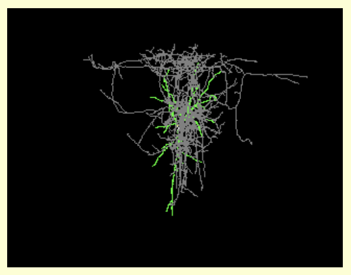
Kravitz Dataset1
The data sets included in this resource are published in Kravitz et al, 2012 Distinct roles for direct and indirect pathway striatal neurons in reinforcement (Nat. Neurosci). The data shows: optogenetic activation of dopamine D1 or D2 receptor-expressing striatal projection neurons influenced reinforcement learning in mice. Stimulating D1 receptor-expressing neurons induced persistent reinforcement, whereas stimulating D2 receptor-expressing neurons induced transient punishment. These 3 recording files contain data collected in 2010 and 2011 by Lex Kravitz in Anatol Kreitzer''s lab at the Gladstone Institutes. Each file contains ~ 1 hour of awake in vivo recording data, containing the spike times for ~30 minutes of spontaneous activity, preceeded or followed by 400 laser pulses (473nm laser light, 1 sec pulses, 3 seconds inter-pulse-interval). The laser pulses were presented at 4 intensities: 0.1mW, 0.3mW, 1.0mW, and 3.0mW, and pulse times at each intensity are given in separate columns in each data file. Finally, each data file contains the identification of ''light-modulated units'' as we identified them (see methods below). Methods: Viral expression of DIO-ChR2-YFP and DIO-YFP We used double-floxed inverted (DIO) constructs to express ChR2-YFP fusions and YFP alone in Cre-expressing neurons, which virtually eliminates recombination in cells that do not express Cre-recombinase (Sohal et al, Nature, 2009). The double-floxed reverse ChR2-YFP or YFP cassette was cloned into a modified version of the pAAV2-MCS vector (Stratagene, La Jolla, CA) carrying the EF-1a promoter and the Woodchuck hepatitis virus posttranscriptional regulatory element (WPRE) to enhance expression. The recombinant AAV vectors were serotyped with AAV1 coat proteins and packaged by the viral vector core at the University of North Carolina. The final viral concentration was 4 x 10^12 virus molecules/mL (by Dot Blot, UNC vector core). This viral construct can now be ordered in single aliquots directly from UNC Vector core as product AAV-EF1a-DIO-hChR2(H134R)-EYFP, at http://genetherapy.unc.edu/services.htm) Implantation of electrode arrays for awake recordings Anaesthesia was induced with a mixture of ketamine and xylazine (100mg ketamine plus 5mg xylazine per kilogram of body weight i.p.) and maintained with isoflurane through a nose cone mounted on a stereotaxic apparatus (Kopf Instruments). The scalp was opened and a hole was drilled in the skull (0.0 to +1.0mm AP, -1.0 to -2.0mm ML from bregma). Two skull screws were implanted in the opposing hemisphere. Dental adhesive (C&B Metabond, Parkell) was used to fix the skull screws in place and coat the surface of the skull. An array of 16 or 32 microwires (35-??m tungsten wires, 100-??m spacing between wires, 200-??m spacing between rows; Innovative Physiology) and one optical fiber in a ferrule was lowered into the striatum (3.0mm below the surface of the brain) and cemented in place with dental acrylic (Ortho-Jet, Lang Dental). After the cement dried, the scalp was sutured shut. Animals were allowed to recover for at least seven days before striatal recordings were made. In vivo electrophysiology Voltage signals from each recording site on the microwire array were band-pass-filtered, such that activity between 150 and 8,000Hz was analysed as spiking activity. This data was amplified, processed and digitally captured using commercial hardware and software (Plexon). Single units were discriminated with principal component analysis (OFFLINE SORTER, Plexon). Two criteria were used to ensure quality of recorded units: (1) recorded units smaller than 100??V (~3 times the noise band) were excluded from further analysis and (2) recorded units in which more than 1% of interspike intervals were shorter than 2ms were excluded from further analysis. Average waveforms were exported with OFFLINE SORTER. During the recording we coupled the array to a laser and pulsed the laser at four intensities (0.1mW, 0.3mW, 1mW, and 3mW). Laser stimulation was run in a cyclical fashion, on for 1 second, and off for 3 seconds. Each neuron received 100 pulses at each laser intensity. Identification of ChR2 expressing units in in vivo recordings For all neurons, peri-event histograms were generated for each laser intensity independently. Neurons were classified as ChR2-expressing if they exhibited a firing rate greater than 3x above the standard deviation of the 1-second preceding the laser pulse within 10msec of the laser onset. Each neuron was tested independently at each laser power, and neurons that satisfied this criteria at any one power were defined as ChR2-expression.
Details
- Resource Type: Resource, data set, data or information resource
- Keywords: dio-chr2-yfp, cre-recombinase, yellow fluorescent protein, channelrhodopsin, optogenetics, striatum, direct, indirect pathway, neostriatum medium spiny neuron, electrophysiology, recording, action potential, multiunit recording, gladstone institute
- Resource ID: SCR_008759
- Proper Citation: (Kravitz Dataset1, RRID:SCR_008759)
- Parent Organization: University of California at San Francisco; California; USA
- Related Condition:
- Funding Agency:
- Relation:
- Reference: DOI:10.1038/nn.3100, PMID:22544310
- Website Status: Last checked down
- Alternate IDs: nlx_144028
- Alternate URLs:
- Old URLs:
- v_uuid: 6df80ca6-d9a1-5c13-a6ca-29f1084498bb


